Local Patients Benefit From PET Scans
- Category: Hospital News
- Posted On:
Positron Emission Tomography (PET) imaging is just one of the advanced tests that Washington Health uses to diagnose, stage, and treat cancer. Most significantly, it can determine whether a tumor has grown or spread, and if radiation and chemotherapy are effective. It provides more information than other types of tests to help doctors develop treatment plans for oncological, cardiac and neurological conditions.
A PET scan creates images of high metabolic activity in the body, rather than pictures of anatomy only, like magnetic resonance imaging (MRI) or computerized tomography (CT scan). Cancer in the body will show up on the PET scan as hyperactive tissue, showing metabolic changes, according to Dusty Finn, the lead certified nuclear medicine technologist at Washington Health. From a PET image, doctors can determine how advanced a tumor is, and how to treat it.
Doctors can also see if treatment is shrinking a tumor, or if the cancer has metastasized or spread. A radiologist will interpret the imaging, and provide a report to the oncologist, who shares it with the patient and develops a treatment plan.
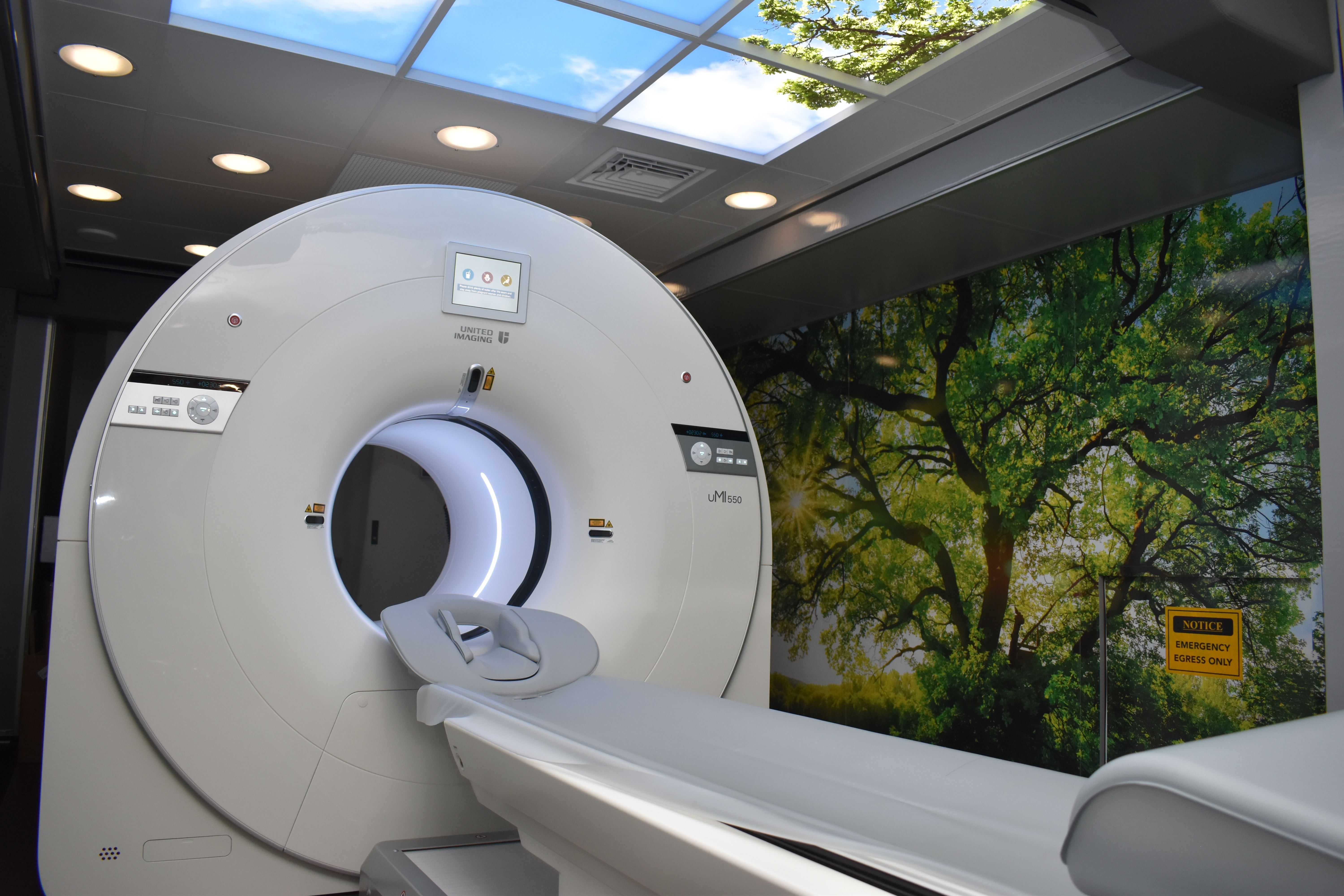
Washington Health has the most advanced type of PET scanner, shortening the time of a scan to about 10 minutes, said Finn. After an IV injection of a radioactive tracer (most are glucose-based agents with no side effects), a patient waits for approximately 45 minutes before being scanned. If they’re wearing clothing with no metal, the scan can be taken in street clothes without changing into a gown.
“We try to make it as easy, convenient and comfortable for a patient as we can,” Finn noted. The images are done in a separate facility across the street from the main hospital.
Promising for Prostate
PET is especially good at detecting prostate cancer, thanks to a radioactive imaging agent that binds to prostate cancer cells and helps doctors identify them. It’s mostly used when prostate cancer returns, or has spread, Finn explained.
Prostate Cancer Awareness Month is observed in September, and a good reminder for men to get their annual prostate screening. Men should talk to their primary care provider about in-office screening.
Cardiac PET to Debut
This fall, Washington Health will offer PET imaging that looks inside the heart to diagnose and help treat some cardiac conditions.
Cardiac PET scanning will allow doctors to check for blocked arteries and blood flow. This test may prevent patients from having to undergo a cardiac catheterization, a minimally invasive procedure that uses catheters and cameras to see inside the heart. PET imaging may also be done before catheterization in some cases.
This new imaging will also benefit people with stents, giving cardiologists insight into their viability and whether blood is flowing well. “Having the Cardiac PET/CT Imaging Program right in Fremont will avoid patients from having to leave the area to go to other health centers for this test.” Finn added.
“We can see myocardial blood flow to check for blockages in persons with chest pain or shortness of breath,” he said. In the past, the only way to see this was through a catheterization, which is a minimally invasive procedure that sometimes requires anesthesia.
Having these images will help diagnose new cases of heart disease, and allow cardiologists to adjust patients’ medications or treatment plans. The new cardiac imaging will employ the same PET/CT scanner used for cancer patients. The only difference will be the IV radio-tracer agent, noted Finn.
Finn and his team are expected to undergo cardiac PET imaging training this summer.
For more information, visit Washington Health’s website at https://www.washingtonhealth.com/services/outpatient-imaging-center/pet-ct-imaging/